“Creeping attachment” has been mentioned in the periodontal literature since the early 1960’s, when the technique of Free Gingival Graft was described as a root coverage procedure.
Up to now, although it’s etiology is unknown, it is believed that is a connective tissue maturation result.
My personal observation is that it’s appearance is not always predictable depending on the harvested area of the palate and the anatomical characteristics of the recipient site additionaly.
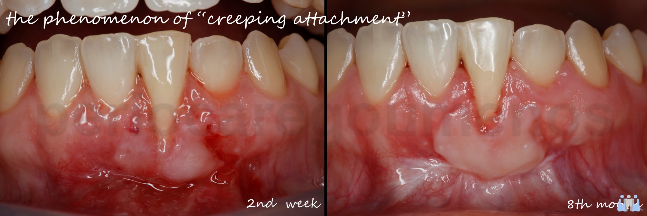
Creeping attachment usually manifests itself either as a coronal migration of the marginal gingiva or as a progressive growth of the soft tissue volume.
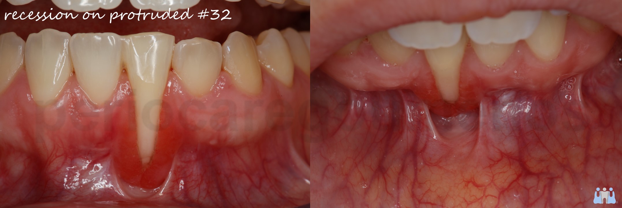
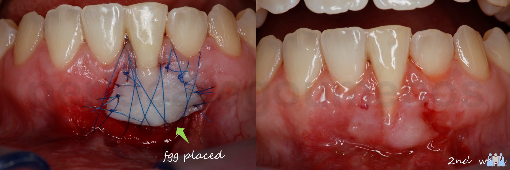
This clinical case presents the 2nd week the root coverage result with the technique of Free Gingival Graft in a very demanding case due to the extension of the recession and the buccal protrusion of the root.
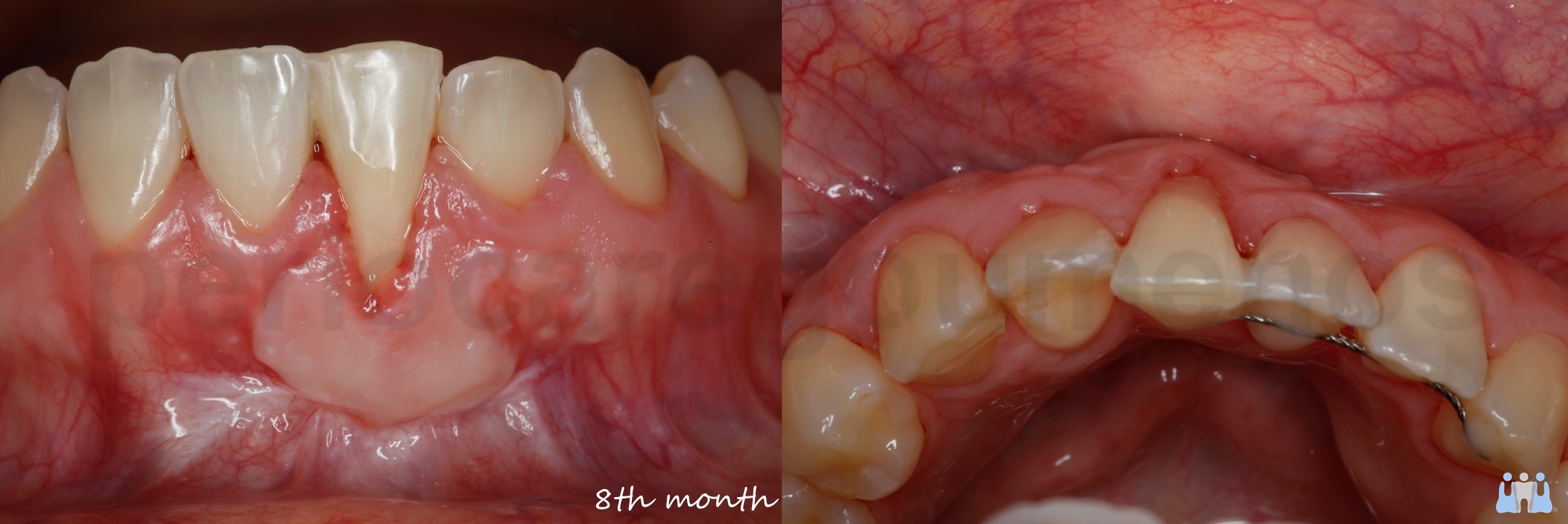
The partial coverage of the root was accompanied with a cleft maybe due to the tension of the protrusion. Although the patient was prepared for a corrective additional procedure based on the 2nd week clinical outcome, the 8-month result revealed surprisingly bridging of the margins of the cleft, improving the clinical result.
Tο φαινόμενο του “creeping attachment” έχει αναφερθεί στη βιβλιογραφία από τη δεκαετία του 60, όταν πρωτοεμφανίσθηκε το Ελεύθερο Ουλικό Μόσχευμα.
Ακόμα και σήμερα δεν γνωρίζουμε την αιτιολογία του συνδυάζοντας το με τη περαιτέρω ωρίμανση του συνδετικού ιστού.
Δύο προσωπικές παρατηρήσεις είναι ότι η εμφάνιση του δεν είναι πάντοτε προβλέψιμη μιας και εξαρτάται από τη περιοχή λήψης της υπερώας, αλλά και από τις ανατομικές ιδιαιτερότητες της περιοχής υποδοχής.
Συνήθως παρατηρείται μια μυλική επέκταση του ουλικού περιγράμματος ή μια προοδευτική αύξηση του όγκου του μαλακών ιστών διαχρονικά.
Στη παρούσα ανάρτηση επιχειρήθηκε κάλυψη της ρίζας με ελεύθερο ουλικό μόσχευμα σε ένα πολύ απαιτητικό περιστατικό, λόγω της έκτασης της υφίζησης αλλά και της παρειακής κλίσης της ρίζας.
Παρατηρώντας το αποτέλεσμα της 2ης μετεγχειρητικής εβδομάδας προετοίμασα την ασθενή για διορθωτική επέμβαση μερικούς μήνες μετά για διόρθωση της σχισμής που είχε εμφανισθεί στο κέντρο της ριζικής επιφάνειας, αποτέλεσμα μάλλον των τάσεων από τη παρειακή απόκλιση της ρίζας, δεδομένου ότι το πάχος των όμορων θηλών ήταν καλό.
Η ασθενής σπουδάζει στο εξωτερικό και εμφανίσθηκε μετά από 8 μήνες.
Το ενδιαφέρον της παρούσας ανάρτησης έγκειται στην όμορη συμπλησίαση των άκρων της σχισμής ως αποτέλεσμα του “creeping”, βελτιώνοντας προς μεγάλη έκπληξη μου σημαντικά τη κλινική εικόνα από αυτή της 2ης μετεγχειρητικής εβδομάδας.